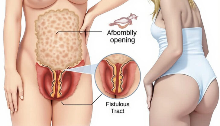
What is Anorectal Malformations (Imperforate Anus)?
Anorectal malformation, often called imperforate anus, is a condition where the individual does not have a normal anal opening. Instead, they have an abnormal passageway, known as a fistulous tract, that opens onto the skin surface near the anus or into the nearby body structures. In males, this tract may connect to the urinary system and in females, it might connect to the reproductive organs.
The severity of the condition often depends on how far the abnormal opening is from the place where the anal opening should be. The further away it is, it’s more likely there will be additional problems, like underdeveloped muscles around the anus. Understanding the exact type of anorectal malformation a person has is extremely important because it can help doctors predict the future health outcomes and decide whether or not the patient will be able to control their bowel movements in the long run.
What Causes Anorectal Malformations (Imperforate Anus)?
The exact cause of anorectal malformations – which are birth defects affecting the area where the rectum, the end of the large intestine, connects to the anus – is unknown. However, there’s a high chance that genetics play a part in causing them. If you already have a child with an anorectal malformation, there’s roughly a 1% chance that your next child will also have this issue.
There are also certain genetic conditions that have higher rates of anorectal malformations. For example, a condition known as the Currarino triad, which is passed down through families, often features these malformations. Similarly, patients with trisomy 21, also known as Down syndrome, frequently have anorectal malformations without a fistula, which is an abnormal connection between organs. In fact, about 95% of patients with Down syndrome also have an anorectal malformation without a fistula, while this is only true for about 5% of all patients with anorectal malformations.
Some evidence suggests that environmental factors may influence the development of anorectal malformations. For instance, there’s a link between these malformations and in vitro fertilization, exposure to a drug called thalidomide, and diabetes. Animal studies also show that exposure to certain chemicals, like trans-retinoic acid and ethylene thiourea, could lead to anorectal malformations.
Risk Factors and Frequency for Anorectal Malformations (Imperforate Anus)
Anorectal malformations are conditions that happen in about 1 in every 5,000 births. These conditions are a little bit more common in boys than girls. Most boys with this malformation have a connection between their rectum and urinary system, known as a recto-urethral fistula. This is true for about 70% of these boys. For girls, the most common type of anorectal malformation is a recto-vestibular fistula, in which there is a connection between the rectum and the vaginal area.

Signs and Symptoms of Anorectal Malformations (Imperforate Anus)
Most newborns with anorectal malformations are identified at birth. It’s important to do a thorough examination and work-up in these babies as around 60% of them often have other anomalies associated with the malformation.
In addition to checking the bottom and anal area, a complete newborn examination should involve listening to the heart for any unusual sounds, examining the limbs for any deformities, and conducting a complete check of the genital and urinary organs.
A normal anus should be in the right location and the right size for the child’s age. For a full-term newborn, the size of the anus should ideally match a 10 to 12 Hegar dilator (a tool used for measuring the size of the anus), and a one-year-old should match about a 15 Hegar dilator. The right location for the anus refers to the anal opening being centrally located within the anal muscle complex. However, this location can’t always be confirmed in a routine clinic visit and the child might need an examination under anesthesia.
The bottom area should be carefully checked for elements like development of the buttocks, a crease or fold in the buttocks, and any sort of opening or hole on the bottom. For girls with an anorectal malformation, a detailed vaginal examination should also be carried out, noting the number of openings on the bottom. These physical examination findings can provide hints about the type of anorectal malformation the child may have.
Testing for Anorectal Malformations (Imperforate Anus)
If a patient is diagnosed with an anorectal malformation, which is a condition where the anus and rectum don’t develop properly, various diagnostic tests are performed. This is important because this condition often comes with other associated abnormalities in about 60% of patients. Specifically, anorectal malformations are often linked with what’s known as VACTERL defects, which involve issues with the vertebrae (the bones in the back), the anus, the heart, the trachea (windpipe), the esophagus (food pipe), the kidneys, and the limbs.
For newborns diagnosed with an anorectal malformation through physical examination, several imaging tests are necessary. After the placement of a tube through the nose or mouth into the stomach, X-rays of the abdomen and chest are taken. These X-rays help check for a condition known as esophageal atresia, where the food pipe doesn’t connect to the stomach, and tracheoesophageal fistula, where the windpipe and the food pipe are connected. They also perform X-rays of the spine to check for any abnormalities of the backbones.
In boys, if there is stool on the skin around the anus but no visible hole or canal (known as a perineal fistula) by 24 hours after birth, a special type of X-ray called an invertogram is done. With this kind of X-ray, babies are positioned with their buttocks raised for at least 15 minutes. This allows any air in the gastrointestinal tract to move to the part of the rectum that is furthest from the anus. This helps doctors see how far up the rectum is located and whether any surgical procedures, like a colostomy (a surgery that brings an end of the colon to the surface of the body), are needed.
In girls with only one visible hole in their perineum (the area between the vulva and the anus) and a diagnosis of cloaca (a condition where the rectum, vagina, and urethra share a common channel), an abdominal ultrasound is performed. This test uses sound waves to create images of the inside of the body. It helps check for an overfilled vagina due to blocked menstrual flow (hydrocolpos) and swelling of the kidneys due to backed-up urine (hydronephrosis).
All patients diagnosed with anorectal malformation should also have an echocardiogram, which is an ultrasound of the heart, to check for any congenital heart issues. Additionally, they should also have a spinal ultrasound, which uses sound waves to create images of the spine, to screen for a condition called tethered spinal cord, where the spinal cord is attached at the base of the spine and cannot move freely. Lastly, doctors calculate a sacral ratio from the X-ray, which provides information about how well the child’s bowel control might develop as they grow.
Treatment Options for Anorectal Malformations (Imperforate Anus)
If a baby is born with a perineal fistula, which is an abnormal connection between the rectum and the skin near the anus, or a rectovestibular fistula (for girls only), where there is an abnormal connection between the rectum and the vagina, a surgery can be done shortly after birth. This is only possible if the baby has no other medical conditions, like a heart defect, that would make anesthesia unsafe. But if the abnormal opening is wide enough, rather than doing surgery right away, doctors can keep the opening from closing by gently stretching it with a medical instrument. This also helps the baby in releasing stools.
In both cases, doctors aim to make sure that the baby is able to pass stool well through the abnormal opening. If they’re managing the condition with dilation (stretching), they typically plan to do surgery to repair it at around three months of age. This is timed to take place before the baby starts solid food, so there’s a lesser chance that constipation could lead to the stretching of the rectum, which may disrupt its function later on. Normally, the surgery to fix these abnormalities doesn’t need to go through the abdomen.
If a baby boy has a urinary fistula, which is an abnormal channel that allows urine to leak into the rectum, doctors will typically do a procedure shortly after his birth to create a stoma, or an artificial opening for waste removal. This procedure allows the baby to grow before undergoing the actual surgery to repair the fistula and helps the baby pass stool through a bowel segment that’s connected to the urinary system. The stoma also allows doctors to conduct a detailed X-Ray to better understand the kind of urinary fistula present, which helps in planning the corrective surgery.
Similarly, if a baby girl is born with a cloaca, being a single opening instead of separate ones for the urinary tract and rectum, doctors will also create a stoma right after birth. Girls with this condition often need more complicated long-term treatment and care plans, and could benefit from a referral to a specialized centre.
The timing of the definitive surgery for more serious abnormalities depends on the exact abnormality and any other associated medical conditions, particularly heart defects. The surgery is generally performed around or after the baby is three months old. It can be done with a single cut at the back if the rectum and the fistula are located below the tailbone level, as shown on a detailed X-Ray. For more complicated abnormalities, like a fistula connecting the rectum and the bladder, laparoscopy, a minimally invasive procedure that uses a camera and small incisions, can be a helpful surgical approach.
What else can Anorectal Malformations (Imperforate Anus) be?
Almost all babies with a perineal opening (an opening near the anus) are diagnosed at birth with an anorectal malformation. However, in some cases, a child could be diagnosed later. This usually happens when the child has a perineal fistula or an anal stenosis – conditions that might go unnoticed during the first few days after birth.
If there is any doubt about the presence of an anorectal malformation, a pediatric surgeon should examine the child while they’re asleep. This will allow them to assess if the anal opening is of the right size and located correctly within the muscles around it. Sometimes, a child might have an anal opening that seems more towards the front than usual, but this can be within normal variation if the opening is the right size and correctly placed within the muscles.
What to expect with Anorectal Malformations (Imperforate Anus)
The future health outcomes for the patients with an anorectal malformation, a birth defect where the anus and rectum do not develop properly, are largely dependent on their long-term potential to control bowel movements. There are three main factors that can help predict this control: the type of anorectal malformation, the sacral ratio, and the quality of the spinal cord.
Firstly, if the malformation (fistula) is far from the normal anatomical location, it reduces the chances of the child gaining control of their bowel movements as they grow older. Secondly, a low sacral ratio (a measure related to the development of the lower part of the spine) could suggest a decreased ability to control bowel movements. Lastly, issues with the spinal cord, like having a tethered cord (a condition where the spinal cord is attached at the base of the spine and lacks free motion), are another negative indicator for future bowel control.
However, even when these factors are present, it’s important to know that a child can still maintain bowel cleanliness and control in social situations. This can be achieved through a suitable bowel management program and by getting the necessary specialized care.
Possible Complications When Diagnosed with Anorectal Malformations (Imperforate Anus)
During surgery, if great care isn’t taken to stay within the correct area of tissue, there can be complications. This might result in mistakenly placing the anus in the wrong location or outside the central area of the anal muscle complex. In men, the urinary structures, including the urethra, seminal vesicles, and vas deferens could get damaged. In women, possible intraoperative complications could include damage to the vagina.
After the operation, problems might also arise. These can include superficial and deep wound infection, a split or gap where the anastomosis was conducted, an outpouching or an increase in the size of the anoplasty, or a narrowing of the anoplasty tube. There’s also a chance of developing recurrent fistulas – abnormal connections – between the urinary system in men or the gynaecological system in women. If the surgical repair is under excessive tension or if the blood supply to the rectum is insufficient, these fistulas could occur. If the urethra or vagina is injured during surgery, it could also result in fistulas. It would be important to provide care to ensure the health of the rectal wall as opposed to the repaired structure, and a fat pad should be put in place to support the repair.
Here is a list of possible complications:
- Incorrect placement of anus
- Damages to urinary structures in men
- Vaginal injury in women
- Superficial or deep wound infection after surgery
- Anastomosis split or gap
- Outpouching or size increase of the anoplasty
- Narrowing of the anoplasty tube
- Recurrent fistulas between the urinary or gynaecological systems
- Injuries to anterior structures, such as urethra or vagina
Preventing Anorectal Malformations (Imperforate Anus)
It is important to instruct parents of a child with a condition called anorectal malformation about potential future problems their child may have with controlling their bowel movements. These problems could persist in the long term. Three factors can help predict these potential continence issues: the type of anorectal malformation the child has, their sacral ratio (a measure of the child’s lower spine), and the condition of their spinal cord.
The type of anorectal malformation is significant because the farther the abnormal opening (fistula) is from its normal place, the less likely the child will be able to control bowel movements. A normal sacral ratio is about 0.9. If a child’s sacral ratio is less than 0.4, it suggests that they may have trouble achieving bowel control.
It’s also crucial to monitor children with anorectal malformations for urinary tract infections. In female children, it is additionally important to keep an eye on the start of their menstrual cycles to ensure no obstructions (Mullerian obstructions) exist. Also, if there’s a spinal defect like a tethered cord (where the spinal cord is attached at the base instead of floating freely as it should), it further reduces the child’s ability to control their bowel movements in the long run.
