
What is Acetabular Fractures (Broken Hip)?
Acetabular fractures, or breaks in the hip socket, mainly happen in young individuals who experience high-speed accidents. However, ever since wearing seatbelts became mandatory, the number of these injuries has dropped significantly to around 3 in 100,000 people. Despite this, there has been a rise in such fractures resulting from falls of less than ten feet, probably due to an increase in conditions causing fragile bones like osteopenia and osteoporosis. Nothing much has changed since the significant 1993 study by Letournel and Judet. Many of their findings about the treatment of these fractures are still considered the best available. Among the key advancements is the development of methods for fixing certain fracture types through a minor puncture known as percutaneous fixation.
Talking a little about the anatomy, the innominate bone, which contributes to forming the hip socket, is created from three bones: the pubis, ischium, and ilium. The top part of the hip socket, where body weight is placed, is known as the weight-bearing dome. The supply of blood to the outer surface comes from four arteries — the superior gluteal, inferior gluteal, obturator, and medial femoral circumflex arteries. Meanwhile, blood to the inner surface is supplied by three other arteries — the fourth lumbar, iliolumbar, and obturator arteries. You can think of the joint surface like an upside-down Y, connected to the joint between the hip and tailbone with a thick piece of bone known as the sciatic buttress. The hip socket is divided into two parts: an anterior and a posterior column. The anterior column has the front half of the iliac wing, extending to the pelvic brim and upper pubic ramus, and it includes the front half of the hip socket’s joint surface. The posterior column starts from the top part of the greater sciatic notch, extends to the greater and lesser sciatic notches, and also includes the ischial tuberosity.
What Causes Acetabular Fractures (Broken Hip)?
Fractures of the acetabulum, or hip socket, often occur due to high impact and can frequently lead to other organ injuries. This type of injury can be serious due to the possibility of damage to the cartilage, which might cause a debilitating condition called osteoarthritis in the future.
There’s a high likelihood of associated injuries in patients with acetabulum fractures. In a large series of cases studied by a researcher named Matta, 50% of the patients had other injuries: 35% had a limb injury, most commonly in the lower limb, 19% had a head injury, 18% had a chest injury, 13% had nerve damage, 8% had an abdominal injury, 6% had genitourinary or urinary tract injuries, and 4% had spine-related injuries.
Even fractures that only involve the acetabulum often require a blood transfusion. In fact, up to 35% of people with isolated acetabulum fractures needed a transfusion in one study.
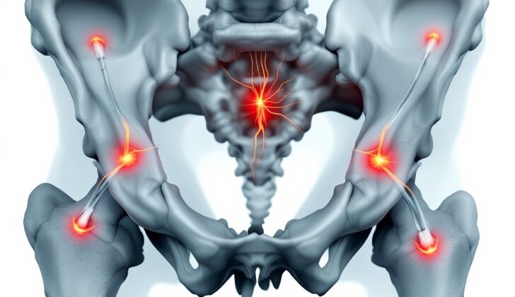
An important part of care after this injury includes checking for damage to the sciatic nerve. The sciatic nerve runs from your lower back, through your hips and buttocks, and down each leg. If this nerve is damaged during the fracture, it frequently affects the part of the nerve known as the peroneal division, which may result in difficulty lifting the foot, a condition known as foot drop. The tibial division of the sciatic nerve is less commonly involved in these injuries.
Risk Factors and Frequency for Acetabular Fractures (Broken Hip)
These injuries frequently happen due to high-speed car accidents, falls from high places, or extreme sports. Over the past few decades, the rate of these injuries has stayed at about 3 for every 100,000 people each year. Similar numbers are reported for fractures resulting from car accidents, but there’s been an increase in fractures from falls that are less than 10 feet. Also, the average age of patients with these fractures has been increasing.
Signs and Symptoms of Acetabular Fractures (Broken Hip)
The first step in assessing trauma is to follow established guidelines for examining and treating injuries. Understanding how the injury happened can guide the treatment plan. The doctor should check the whole body for other injury signs. It’s also important to thoroughly check the musculoskeletal system, mainly the nerves in the arms and legs, as well as the skin. Special attention should be given to the soft tissue to detect possible Morel-Lavalle lesions, a type of injury that affects the skin and underlying fat tissue.
Testing for Acetabular Fractures (Broken Hip)
In order to diagnose a fracture in the acetabulum (the part of your pelvis that meets your hip), a plain X-ray might be used initially. This scan can show quite a bit, but if you have injuries in multiple places, a CT scan may be necessary because they can provide a more detailed image. The physician might order both a standard X-ray as well as specialized views known as Judet views, which focus on the pelvis area. Identifying certain landmarks or structures in these images can help classify the fracture.
In the X-rays, physicians look for the following landmarks: iliopectineal line, ilioischial line, teardrop, roof of acetabulum, anterior wall, and posterior wall. These landmarks are not actual anatomical structures but areas that help visualize and understand the fracture better. For instance, the two lines represent the anterior and posterior columns of the pelvis, while the teardrop represents an area used to identify certain features of the fracture.
CT scans have made it easier to diagnose and classify fractures in the acetabulum. These types of scans can provide precise information about the fracture such as location, presence of any fragments within the joint, and the direction of the fracture lines. For physicians who are less experienced, 3D imaging can be helpful. There’s also a method called dynamic stress X-rays, which helps evaluate the stability of the hip joint, particularly crucial when there’s a specific kind of fracture involving the posterior wall. If your hip displaces during this exam, that indicates instability in the joint.
The Letournel system is commonly used to classify acetabulum fractures. They are divided into five elementary types and five associated types. Each type involves a different area and features of the acetabulum and is associated with different indicators on the X-ray or obturator oblique image. Some types are more common than others, and the different patterns of fractures can give a clue as to the type and severity of the injury. For example, a transverse fracture is a type where both the front and back columns of the acetabulum are broken, which is noticeable as a disruption in both the ilioischial and iliopectineal lines on an AP view.

Treatment Options for Acetabular Fractures (Broken Hip)
Open reduction and internal fixation are procedures often required to heal fractures in the acetabulum, the part of the pelvis that forms the hip joint. However, some individuals might not need these procedures. Examples could include fractures that are stable and haven’t shifted, those that don’t involve the dome of the acetabulum (the upper weight-bearing part of the socket), or those that are low on the anterior column (the front part). In these cases, non-surgical management might be an option.
A CT scan can help doctors determine if a fracture can be managed non-surgically. By evaluating the ‘vertex’, the tip of the acetabulum, they can assess the extent of the injury. If the area around the vertex is not fractured, and the shifted bone does not exceed 2mm, non-surgical management may be suitable.
For those managed without surgery, the initial step is bed rest to help manage the pain. Once the pain is bearable, the patient can start moving with limited weight-bearing, which is gradually increased over 6 to 12 weeks as the fracture heals. However, If surgery is indicated but not possible due to the patient’s health conditions, extended bed rest may be necessary.
Surgical repair is typically needed when the fracture destabilizes the hip joint, when fragments of bone or tissue are trapped in the joint, or when non-surgical management is not practical. In these instances, a total hip replacement or percutaneous fixation (a minimally invasive procedure) might be considered.
Percutaneous fixation is becoming a popular technique, especially for patients who are too ill for extensive surgery or as a supplement to open reduction and internal fixation. This procedure involves inserting a guidewire into the fractured area to guide the placement of screws. The exact method varies depending on the column of the acetabulum that is fractured.
Timing for surgery is usually 3 to 5 days after the injury as delays over 3 weeks could lead to worse outcomes. Emergency surgery may be required in certain cases like a repeating hip dislocation, nerve damage, blood vessel injury, or an open fracture.
Deciding which surgical technique to use depends on the type and location of the fracture, how long it’s been since the injury, and where the largest displacement of the bone is located. After the fracture is positioned correctly and stabilized, plates are added for additional support. Healthy, young bone may be stabilized using screws alone; however, weakened bone and all wall fractures usually require additional support with plates.
What else can Acetabular Fractures (Broken Hip) be?
It’s important not to mix up fractures of the acetabulum (the part of your hip socket) with other types of fractures. Thanks to computed tomography (CT) scans, we can better identify these differences. But we shouldn’t overlook other possible fractures such as:
- Fractures of the pelvic ring
- Fractures of the upper part of the thigh bone.
Care should be taken especially in older adults who suffer fractures from minor falls; in these cases, it’s very important not to miss what might be a pathological fracture (a break in a bone weakened by some other disease).
What to expect with Acetabular Fractures (Broken Hip)
When it comes to fractures to the acetabulum (the part of your hip bone that forms the hip socket), they have typically had a less favorable outlook due to the high impact of the injury and accompanying injuries. However, with modern-day techniques like open reduction (resetting or realigning the bone fragments through surgery) and internal fixation (stabilizing the fracture by attaching metal plates or inserting screws or nails into the bone), the outlook for these kinds of fractures is now mostly positive.
Still, the exact outlook depends on various factors, such as the pattern of the fracture (a “T-type” fracture generally has the worst outlook), the state of the hip at the time of injury (including femoral head lesions, changes in the margin of the hip joint, signs of a ‘gull sign’ which suggests a particular type of damage, the extent to which the hip joint has been successfully realigned, and the stability of the joint after treatment).
The most reliable way to predict the result after treatment is the condition of the hip and the X-ray images after a year, as most hip conditions don’t improve after that period.
Possible Complications When Diagnosed with Acetabular Fractures (Broken Hip)
Acetabular fractures can lead to some complications. These could include:
- Post-traumatic arthritis and bone death (osteonecrosis); The quality of the fracture reduction plays a major role in the risk of later arthritis. The aim is to achieve a reduction within 1 millimeter.
- Infection, which occurs in about 5% of cases.
- Accidental injury to nerves during treatment (Iatrogenic nerve injury).
- Blood clot in the veins (4% symptomatic DVT), and pulmonary embolism (1% PE)
- Medical hardware left in the joint (Intra-articular hardware).
- Abnormal growth of bone in the muscle tissue (Heterotopic ossification).
Recovery from Acetabular Fractures (Broken Hip)
Before undergoing the procedure, patients who are in skeletal traction (a method of using weights and pulleys to align broken bones) can experience changes in their skin bacteria since they can’t clean their perineum (the area between the anus and the genitals). Therefore, preventative antibiotics are usually recommended, which often includes a type of antibiotic called a first-generation cephalosporin and some additional coverage against certain bacteria (gram-negative). Drains are typically utilized for about 48 hours or until there’s no more fluid drainage.
There’s a high risk of developing blood clots in the leg veins, also known as deep vein thrombosis (DVT). To reduce this risk, mechanical compression devices are used on both legs from the time of admission until the patient is ready to go home. Furthermore, medication to prevent blood clots is administered until the patient can move around (usually for about 6 to 12 weeks).
For surgeries that involve extended or posterior approaches, there may be a risk of a condition called heterotopic ossification – where bone tissue forms in places where it shouldn’t. The prevention of this condition can include a single dose of radiation (700 to 800 cGy) within 72 hours after surgery, or a medication called indomethacin (25 mg three times daily) starting within one day after surgery and continued for six weeks. However, the use of indomethacin is not advised for fractures at high risk of not healing properly.
The patient is usually advised to place minimal or no weight on the affected side for 2 to 3 months after surgery, then allowed to put weight on it as tolerated. Physical therapy typically begins the day after the operation.
Preventing Acetabular Fractures (Broken Hip)
Preventing fractures in your hip socket, or acetabulum, can be challenging, however, always wearing a seatbelt when driving can reduce the risk. For older individuals, screenings for weakened bones, also known as osteopenia or osteoporosis, are beneficial. DEXA scans, which are a type of x-ray that measures bone density, and treatments that include Vitamin D or a type of medication called bisphosphonates can aid in prevention. Additionally, taking steps to prevent accidental trips or falls can also help in avoiding brittle bone fractures.